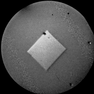
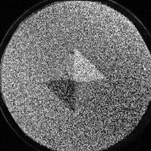
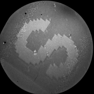
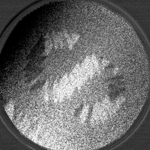
Last week, the installation of the PEEM photoelectric microscope on the PEEM / XAS experimental line was successfully completed. For the first time at a Polish synchrotron, synchrotron radiation was used to obtain imaging with chemical and magnetic resolving power. As a result of intensive XAS spectroscopic measurements, several experimental samples were tested. The most spectacular was the structured permalloy sample on a silicon substrate with a Co spacer covered with a protective layer of aluminum (1nm Al / 2nm NiFe / 20nm Co / Si). On the surface of the sample there were various nano- and microstructures obtained by the lithographic method, which showed a rich magnetic domain structure.
S for Solaris. The above magnetic images were collected on the iron L3 line for a radiation energy of 706.8eV. On the left, an XAS image acquired with linear polarization. On the right, the XMCD differential image of the magnetic domains. The field of view is 75μm.